15 years experience with extracellular vesicles
Our group's expertise covers a variety of isolation techniques, functional assays and molecular testing.
 Contact
 University of Szeged · Department of Immunology
From fundamental vesicle biology to reliable biomarkers and therapeutic insight.
Who we are
The Extracellular Vesicle Research Group, led by Dr Krisztina Buzás, studies how cell-derived vesicles carry information in health and disease.
By combining immunology, proteomics, advanced imaging and data analysis, we aim to translate basic vesicle biology into reliable biomarkers and therapeutic insight. Our interdisciplinary team works with clinicians, engineers and informatics experts to turn discoveries into practical benefit.
Our group's expertise covers a variety of isolation techniques, functional assays and molecular testing.
We conduct in vitro assays with primary and tumour cell lines, including wound healing, migration, spheroid models and perfusion systems such as MIVO®.
We work with mouse melanoma, disease-induction and air-pouch models, integrating them safely into extracellular-vesicle experiments.
Our work combines advanced statistical methods with supervised and unsupervised machine-learning techniques.
Ongoing projects
We study vesicles from molecular composition to multicellular communication. Select a topic to explore the study and open its figure at full size.
Vesicle trafficking
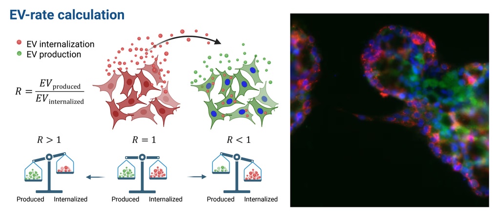
We quantitatively characterize the dynamics of extracellular vesicle (sEV) trafficking in the tumor microenvironment by measuring both vesicle release and uptake across multiple cell types, including different tumor, and stromal cells. Using dual-color in-cell EV labeling combined with flow cytometry, we can simultaneously track sEV production and internalization without isolation, thus preserving physiologically relevant concentrations. This allows us to calculate the EV-dynamic profile, a robust metric expressing the balance between vesicle release and uptake for each cell type. By applying this method under baseline and stress conditions, we identify cell type–specific communication patterns and, importantly, can monitor shifts in vesicle exchange during treatments — capturing not only changes in release but the full dynamic balance of vesicle trafficking.
 View full-size figure
 Immunomodulation
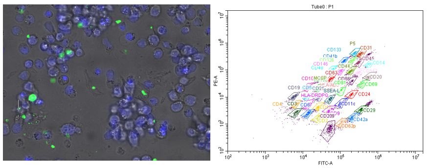
Extracellular vesicles carry a diverse molecular cargo that plays a pivotal role in immunomodulation, emerging as key regulators of immune responses during tumor progression. Tumor-derived EVs often harbor immunosuppressive factors that impair the antitumor activity of immune cells. In this study, we aim to investigate the immunological effects of blood-derived small EVs on B cells from different brain tumor patients. Since brain tumor-derived EVs primarily interact with microglia, we further examined their immunomodulatory properties in a microglia–B cell co-culture system, focusing on vesicle-driven alterations in cytokine and chemokine secretion, modulation of immune checkpoint proteins (PD-1, CTLA-4) and surface marker expression, as well as their influence on B cell proliferation and apoptosis.
 View full-size figure
 3D culture · MIVO®
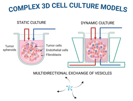
To capture the dynamic interplay of extracellular vesicles (sEVs) within the tumor microenvironment, we employ complex 3D cell culture models that recapitulate key structural and functional features of in vivo tumor tissues. These models are operated under two distinct conditions: a static culture with steady medium, allowing controlled baseline observations, and a dynamic flow system (MIVO®) that better mimics physiological fluid circulation in the human body. The inclusion of dynamic flow enables more accurate modeling of vesicle trafficking and exchange between tumor, fibroblast, endothelial, and immune cells. By integrating advanced imaging with quantitative vesicle tracking, we map the multidirectional exchange of vesicles across different cell populations, revealing how sEV communication networks orchestrate tumor progression, immune modulation, and fibroblast remodeling. This dual-condition approach provides a versatile platform for dissecting cell–cell communication at high resolution and for evaluating therapeutic strategies aimed at disrupting pathogenic vesicle signaling pathways.
 View full-size figure
 Molecular fingerprinting
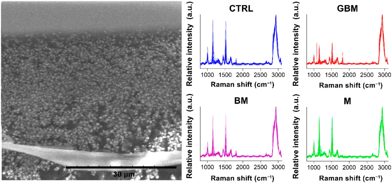
The vesicle research community, including our research group, posits that it may be more appropriate to consider the vesicle as the fundamental unit of information, rather than focusing on the molecular contents of vesicles through conventional omics approaches. In this context, Raman spectroscopy — a type of vibrational spectroscopy — offers a compelling alternative, as it enables the characterization of the complete molecular profile of a biological sample. Our group employs this molecular fingerprint to facilitate the diagnosis of various tumor types, including central nervous system tumors, through the analysis of blood-derived extracellular vesicles.
 View full-size figure
 Proteomics · WGCNA
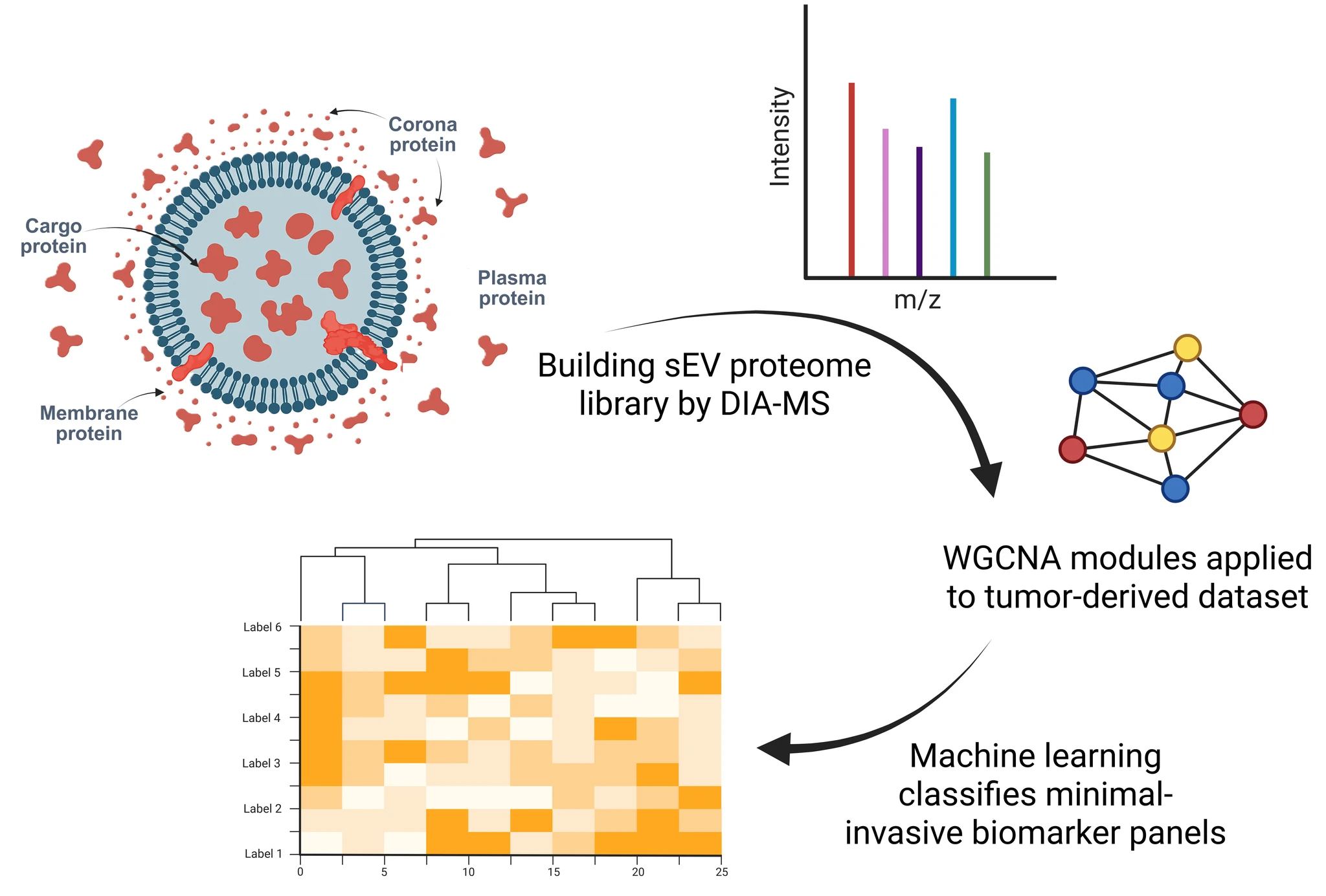
We are building a high-coverage EV-proteome library by deep DIA-LC-MS profiling of plasma EVs from healthy volunteers. Quantitative intensity tables are processed with Weighted Gene Co-expression Network Analysis (WGCNA) to translate thousands of peptides into a biologically interpretable surface protein atlas. Applying the same network to tumor-derived datasets highlights the concerted protein-expression changes that distinguish CNS-tumor EVs from healthy controls and nominates minimal-invasive biomarker panels. Combined with machine-learning classifiers, this atlas-driven workflow offers a reproducible, expandable pipeline for diagnostic discovery and for monitoring therapeutic response in future multicenter cohorts.
 View full-size figure
 Protein corona · Immunology
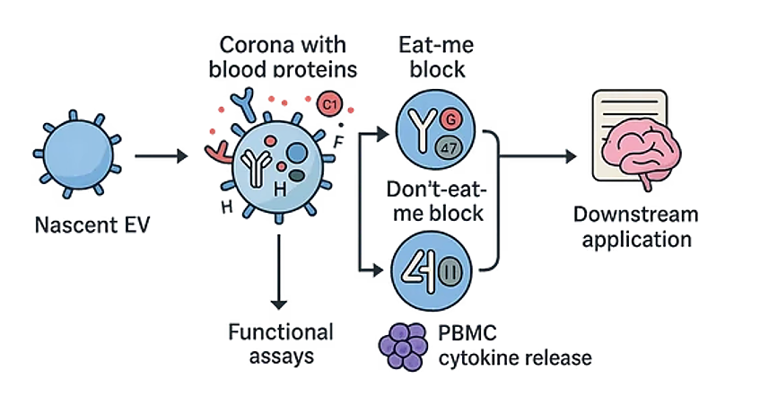
Our group aims to unravel how the blood-derived protein corona dictates the immunological fate of circulating extracellular vesicles. Using nascent endothelial sEVs as a model, we map the balance between “eat-me” (IgG / C1q) and “don’t-eat-me” (CD47 / factor H) corona blocks and link these signatures to functional read-outs—complement activation, macrophage uptake and PBMC cytokine release. This growing atlas will serve as a reference for forthcoming comparisons with glioblastoma plasma and already highlights surface modules that can be exploited as biomarkers or therapeutic targets to overcome tumour-EV immune evasion.
 View full-size figure
 Interested in one of these projects?
Start a conversationOur group
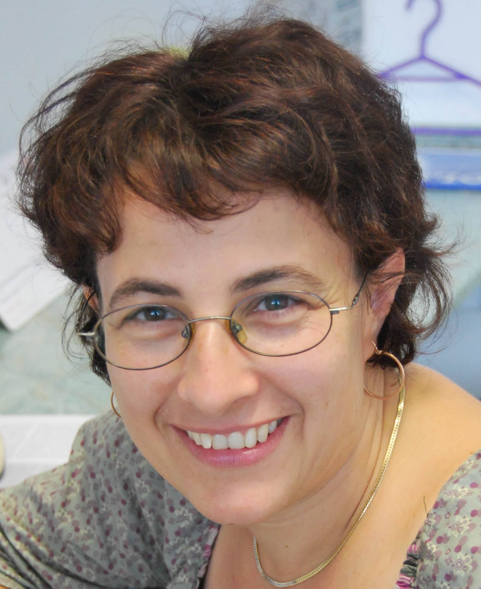
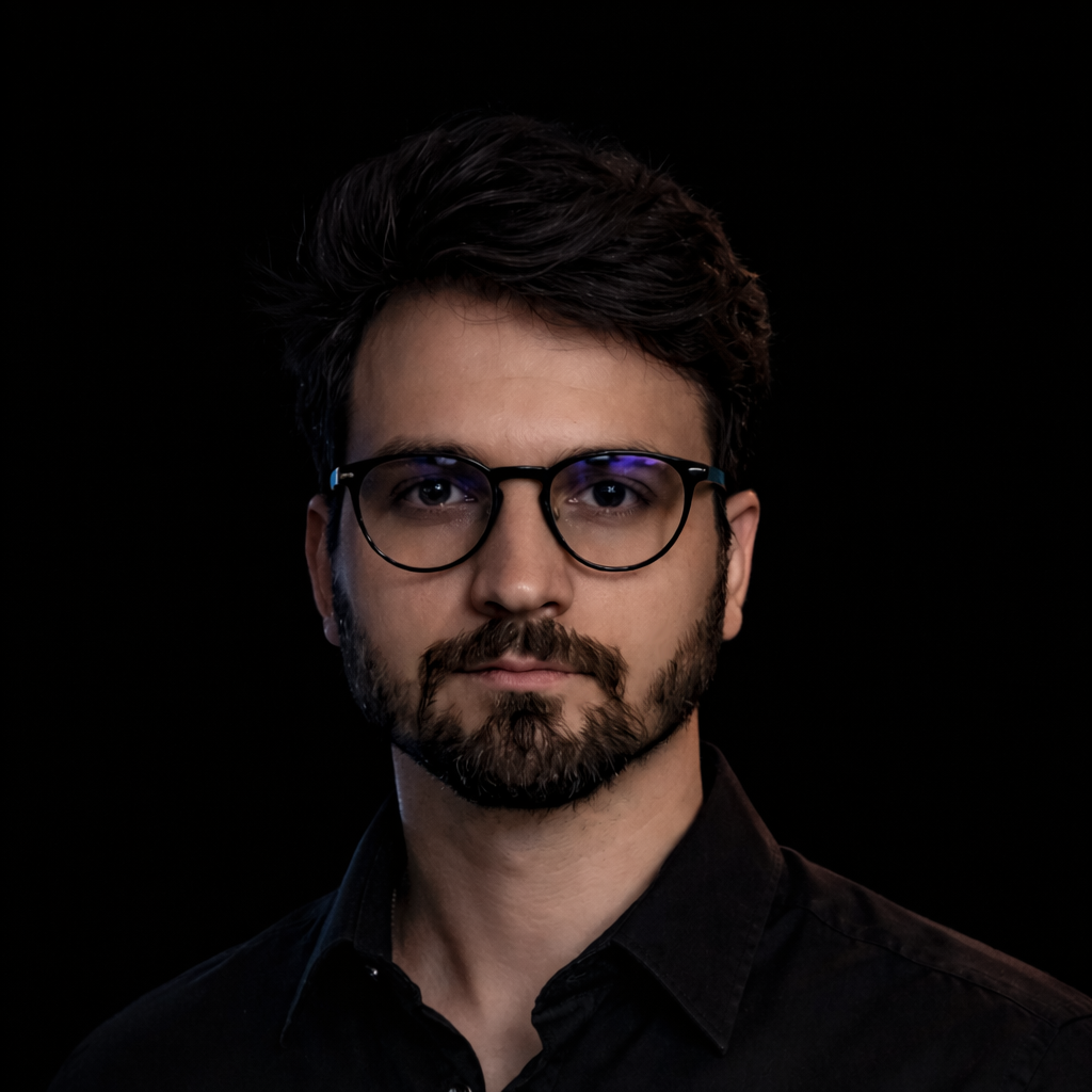
Our expertise spans molecular biology, clinical research, data analysis and bioinformatics. The strength of the group lies in how these disciplines connect.




Alumni


Selected publications
Peer-reviewed work from extracellular-vesicle standards and proteomics to tumour models, machine learning and clinical biomarker discovery.
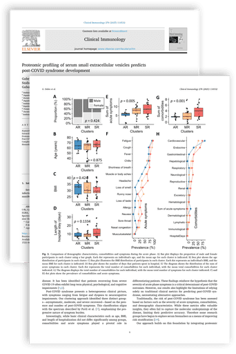
Mass-spectrometry profiling of serum sEVs from 59 stratified patients identified vesicular protein signatures that separated future post-COVID cases from fully recovering individuals at hospital admission. Innate-immune and coagulation-related proteins emerged as strong early predictors.
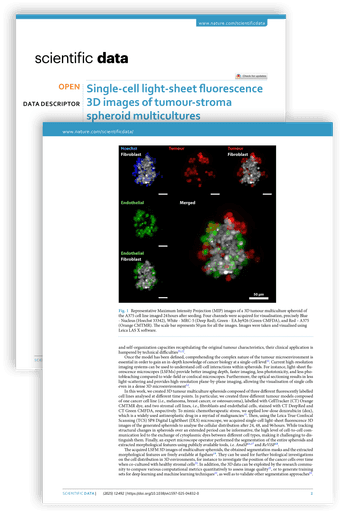
An open, high-resolution three-dimensional image atlas of tumour–stroma spheroids captures spatial heterogeneity and cell interactions at single-cell level, supporting image-analysis development, drug-penetration studies and more realistic in-vitro models.
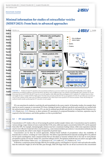
The international MISEV 2023 task force updates experimental and reporting standards for EV isolation, characterisation, cargo analysis and functional assays, with new guidance for in-vivo tracking, single-EV technologies and clinical-grade preparations.
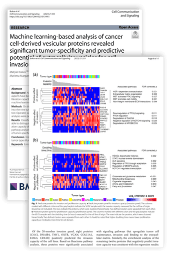
A machine-learning meta-analysis of more than 3,700 EV proteins uncovered signatures that distinguished tumour types with 90% accuracy and identified protein subsets associated with aggressive behaviour.
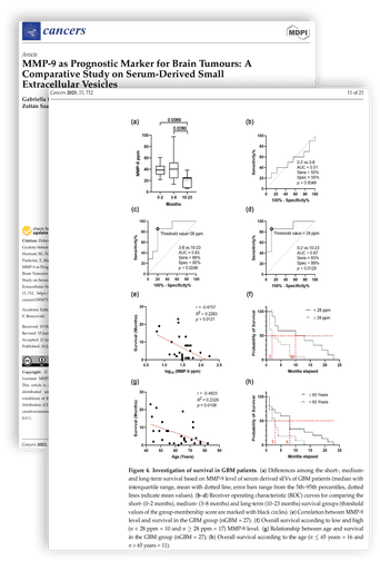
A comparative study of serum-derived small extracellular vesicles evaluating MMP-9 as a prognostic marker across brain tumours.
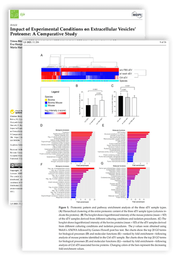
Subtle changes in culture media, isolation kits or storage temperatures can reshape EV protein profiles, alter pathway-enrichment results and introduce artefacts. The study provides standardisation guidance and a reference dataset.
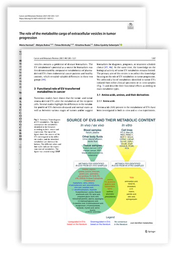
A synthesis of 95 studies catalogues more than 100 metabolites enriched in tumour-derived EVs and highlights their roles in cancer-cell fuelling, microenvironment remodelling and immune suppression.
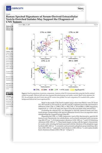
Label-free Raman spectroscopy on 138 patient sera identified distinct fingerprints for glioblastoma, brain-metastatic lung cancer and meningioma, supporting rapid EV-based neuro-oncological diagnostics.
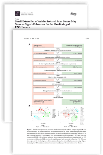
sEV fractions concentrated 65 tumour-related proteins while depleting 129 abundant serum components, producing clearer molecular contrasts between CNS tumour entities than whole serum.
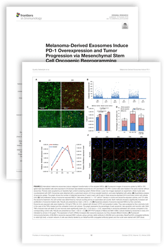
Melanoma exosomes converted naïve mesenchymal stem cells into PD-1-high, melanoma-like cells that foster tumour growth, revealing a mechanism of oncogenic rewiring, immune evasion and metastasis.
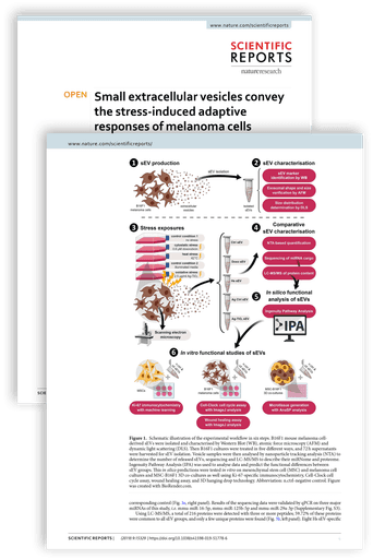
Exosomes released under oxidative, heat or cytostatic stress carried stress-specific cargo that increased proliferation, migration and three-dimensional micro-tissue formation in recipient cells.
Methodological toolbox
Open a method group to see the techniques, purpose and key instrumentation used in our work.
Highly defined vesicle population produced under controlled in vitro conditions.
Tissue culture flasks & bioreactorsSystemic snapshot of circulating EVs, collected with anticoagulant to minimise clotting artefacts.
EDTA or sodium-citrate VacutainerPlatelet-enriched EV mix released during coagulation; convenient for routine clinical sampling.
Serum-separator tubeLow-background matrix for neuronal and glial EVs; limited volume but high specificity.
Polypropylene CSF tube (lumbar puncture)Non-invasive access to nephron- and urothelial-derived EVs; osmolality-dependent yield.
Sterile mid-stream urine cupComplex mix of host- and microbiota-derived EVs reflecting the gut microenvironment; requires additional clean-up steps.
Stool collection kitClassic density-based separation, suitable for large sample volumes.
T-1270, Thermo Fisher ScientificGentle, size-based purification for high-purity, intact vesicles.
qEV, IZONMeasures vesicle-size distribution and concentration.
NanoSight NS300, MalvernHigh-resolution analysis of individual particle size, concentration and charge.
Exoid, Izon ScienceProvides high-resolution images of vesicle morphology.
JEM-1400 Flash, JEOLHigh-resolution surface topography and mechanical properties.
Asylum Research, Oxford InstrumentsDetects specific protein markers to confirm vesicle identity.
XCell SureLock, Thermo FisherHigh-throughput analysis of surface markers on single vesicles.
CytoFLEX S, BeckmanLabel-free chemical fingerprinting of vesicle composition.
SENTERRA, BrukerComprehensive proteomic and lipidomic profiling.
Orbitrap, Thermo FisherHigh-resolution 3D imaging of cells and vesicle trafficking.
VisiScope, VWR; SP8, LeicaAutomated cell imaging for large-scale experiments.
Operetta, PerkinElmerFrequentist and Bayesian approaches, including experimental design and teaching.
JASP, Jamovi, IBM SPSS, GraphPadSupervised and unsupervised methods for pattern recognition and predictive modelling.
Orange Data Mining, R, PythonCollaboration network
Our collaborators connect basic science, clinical expertise, imaging, computation and engineering across six institutions.
Contact
For scientific collaboration, doctoral opportunities or institutional enquiries, contact the relevant location directly.
Department
Faculty of Medicine, Department of Immunology
Research laboratory
Institute of Biochemistry